Human brain organoid research sheds light on neurological development and disease
Guest Author: Jennifer MacArthur
Dissecting models of early brain development with single cell gene expression.
The human brain is immensely complex and challenging to research. Processes unique to human brain development cannot be studied in model organisms, and are not experimentally accessible to study in vivo. New techniques to grow functional human brain tissue in vitro are helping neuroscientists and developmental biologists observe and analyze aspects of early brain development that they couldn’t before. We are excited to see 10x Genomics technology being used with these model systems to gain insights about human neurological development and disease at the molecular and cellular level.
Two recent papers utilized human brain organoids and single cell gene expression to study cortical development. Brain organoids are tiny, lab-grown models of developing brain tissue. Scientists make these “mini-brains” starting with human embryonic stem cells or human induced pluripotent stem cells, then coaxing these to become neural progenitor cells. Under the right culture conditions, the neural progenitor cells self-assemble into three-dimensional structures, and mature to form cell types and organization that resemble human embryonic brain development. To model neurological disease, researchers can also create isogenic brain organoids from a patient’s skin or blood cells. Single cell gene expression allows brain development researchers to determine the precise identity and fate specification of diverse cell types for thousands of cells at once, and track organoid development at fundamental resolution.
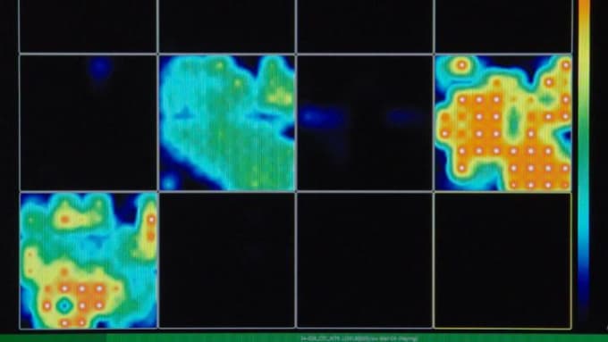
One study, led by Alysson Muotri at University of California San Diego, was able to work out human brain organoid culture conditions that encourage formation of active neural networks. Single cell gene expression revealed the dynamics and details of cell populations as organoid tissue matured. Tracking the transition from progenitor cells to specific neuronal and glial populations showed the cellular basis for neural network activity. They found that when both inhibitory and excitatory neurons had developed, the cortical organoids formed small-scale networks with synchronized electrical spikes and complex brain waves. The scientists hope to optimize this organoid protocol to study effects of drugs and genes on diseases associated with neural network malfunctioning, such as autism, epilepsy, and schizophrenia (1).

Another study, from the laboratory of Gray Camp at Max Planck Institute, created brain organoids to functionally test theories of human brain evolution. The authors generated cerebral organoids from human stem cells, and from chimpanzee and macaque stem cells, then tracked dynamics of gene regulation at single cell resolution. Because protein coding sequences are highly conserved among great apes, the differences between primate species are primarily related to gene expression. Interestingly, the chimpanzee or macaque mini-brains matured more quickly than the human organoids Looking at single cell transcriptomes and chromatin accessibility profiles enabled mapping of cell types and states to determine human-specific features of forebrain development, and pinpointed candidate genes for further research (2).
Ali Brivanlou and colleagues at The Rockefeller University in New York used a different human brain tissue technique to model the earliest stages of nervous system development. They made self-organizing neuroloids from human stem cells and determined single cell gene expression profiles of lineages involved in neural tube formation. To research developmental aspects of neurodegenerative disorders, they also created isogenic neuruloids from Huntington’s disease patients. Surprisingly, in these mutant neuruloids, the three-dimensional tissue structure collapsed. Single cell gene expression identified differentially expressed genes, and pointed to problems with actin-associated tissue organization. Even though the first signs of Huntington’s neurodegeneration do not typically arise until middle age, this experimental model could provide more clues to the origins of Huntington’s disease (3).
These brain organoid and neuruloid experiments demonstrate the importance of recreating a more physiological embryonic context to replicate the natural interplay between different cell types. Single cell gene expression enables researchers to comprehensively characterize the cellular dynamics of development in these models. 10x Genomics solutions offer the resolution and scale to track the molecular changes that drive development and are dysfunctional in neurologic disease. Studying these developmental models with transcriptomic detail can provide insight into possible therapeutic interventions that advance human health.
Explore our solutions for neuroscience research →
Learn more about the Chromium Single Cell Gene Expression Solution →
References
-
C Trujillo et al., Complex Oscillatory Waves Emerging From Cortical Organoids Model Early Human Brain Network Development. Cell Stem Cell. 25, 1–12 (2019).
-
S Kanton et al., Organoid Single-Cell Genomic Atlas Uncovers Human-Specific Features of Brain Development. Nature. 574, 418–422 (2019).
-
T Haremaki et al., Self-Organizing Neuruloids Model Developmental Aspects of Huntington's Disease in the Ectodermal Compartment. Nat Biotechnol. 37, 1198–1208 (2019).
