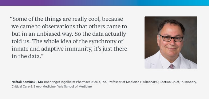
From pulmonary fibrosis to COVID-19: how single cell atlases are advancing our understanding of lung disease
Idiopathic pulmonary fibrosis (IPF) is a devastating lung disease without a cure. Dr. Naftali Kaminski, Professor of Medicine (Pulmonary) at Yale School of Medicine, is one of the trailblazers that has been advancing our understanding of the molecular and cellular basis of the disease. Using single cell sequencing, his laboratory has built an IPF Cell Atlas that is helping define disease-specific transcriptional profiles and functions in disease pathophysiology.

On any given day, you take upwards of 17,000 breaths without much effort. However, for someone living with chronic lung disease, such as idiopathic pulmonary fibrosis (IPF), every breath becomes precious. As the name suggests, pinpointing the cause or origin of IPF, a progressive and fatal lung disease characterized by irreversible scarring of the distal lung, has proven elusive. Dr. Naftali Kaminski of the Yale School of Medicine has devoted his career to understanding the cellular and molecular mechanisms underlying the disease and building support structures for individuals living with this debilitating disease.
A physician by training, Dr. Kaminski completed a pulmonary medicine fellowship at Sheba Medical Center in Tel-Hashomer, Israel, after residency. He then moved to the laboratory of Dean Sheppard at the University of California, San Francisco, where, “...there was some serendipity that I became the person—sort of the first person ever—to do transcriptomic analysis of human fibrotic lungs. This was the early days of microarrays. Now we measure hundreds of thousands of cells and tens of thousands of genes. Then, doing 500 genes at the same time was a big deal.”
In those early days of his IPF research, genomic data from techniques such as microarrays helped uncover the molecular processes unfolding in the lungs during disease progression. “At the time, the notion was that this lung was basically a dead sac of tissue—there's nothing you can do—but now we start looking at the genomics, we start looking at other things, so it's actually an active process,” he said. “In some ways, the word ‘scarring’ of the lungs—‘Fibrosis’—is a little bit of a misnomer. What we see is remodeling of the lung. The lung is actively repairing, which is destroying its function.”
Beyond wanting to advance the scientific and medical understanding of the disease, Dr. Kaminski also became driven to improve the quality of life for patients. “After I published one or two of these papers, I became the director of the Simmons Center [for Interstitial Lung Disease at the University of Pittsburgh] and suddenly started to know the patient population. To some extent, this was, for me, another revelation. There was the feeling that these patients were invisible. At the time, there were no support groups. There was very little public attention. Again, the therapies were not good.”
Going beyond the tissue and into single cells
In recent years, Dr. Kaminski and his laboratory have continued to magnify our understanding of idiopathic pulmonary fibrosis by creating a detailed atlas of lung cell populations to dissect their role in the disease. Despite the advancements that had already been made from studying human fibrotic tissue, he noted that moving to single cell resolution was an important step because “as somebody who did tissue profiling for a living for many years—and actually used a lot of bulk tissue with validation—I’ve always been aware of the sort of hidden ignorance that was in our work.” Furthermore, despite IPF being a “whole organ” disease, Dr. Kaminski realized that to go beyond treatments that only slow progression and find a way to “reverse engineer or reprogram the organs, you need to understand signaling at the single cell level.”
Taking advantage of single cell RNA-sequencing, the IPF Cell Atlas, developed in collaboration with Ivan Rosas, profiled 312,928 cells collected from the lungs of human IPF patients, chronic obstructive pulmonary disease patients, and healthy donors as controls (2). The transcriptomic analysis revealed increased proportions of airway epithelial cells in IPF lungs and, even more intriguing, profound gene expression changes in almost every epithelial cell type. One particular population of these cells, termed aberrant basaloid cells, exhibited a previously undescribed transcriptional signature featuring high expression levels of genes implicated in the pathogenesis of IPF and was marked by features characteristic of cells involved in distal airway development and repair.
Thinking back on the study, he noted that, “This [single cell] technology is really powerful. I started my career in microarrays, and the biggest fear with microarrays was that nothing was reproducible. It took us a while to learn how to normalize the data to get rid of the noise. Single cell is actually really young, but we find extremely consistent signals all the time and in relatively small numbers of samples.” This reproducibility is evident with corroborating single cell sequencing data from Habermann et al (3).
The researchers in the Kaminski laboratory are not the only ones benefiting from the IPF Cell Atlas. One of the project goals was to make the data publicly available in a way that made it valuable to other researchers, and they have managed to do so successfully. “So if you look at the IPF Cell Atlas, I think we now have 7,000 people that have used it. It's almost like you could say that it’s a disease awareness program...just because people who go there would never have been interested in IPF,” he observed. “When we looked at the searches initially, it was really fibrosis genes, but with the pandemic, ACE2 is now the number one searched gene in our atlas. So...these single cell resources and repositories, if they're done in a way that allows people who are not computational biologists to look at them, they become a really useful tool for the community.”
Read more about IPF Cell Atlas or explore it on the Data Mining portal.
Contributing to our understanding of COVID immunopathology
Although Dr. Kaminski has a long-standing research history in pulmonary fibrosis, he also investigates other lung diseases. Most recently, he joined forces with other researchers from Yale University, Howard Hughes Medical Institute, and Shanghai Jiao Tong University to profile innate and adaptive immune system responses in COVID using a single cell multiomics approach (4).
Performing such a comprehensive, multimodal analysis in an ongoing pandemic was no small feat. “You know, I’ve done big data and big science type of work, and this was really special. The two first authors are the people who drove it, but so many of us did equal amounts of work that it's been very unique,” said Kaminski. “When the epidemic just started, there was a group of Yale investigators, like Albert Ko, Akiko Iwasaki, Ruth Montgomery, Charles Dela Cruz, and a few others that said, ‘Ok, we need to have a tissue bank upfront.’”
The study, currently available in preprint on bioRxiv, profiled samples from patients with stable or progressive COVID-19. Cells from both stable and advanced cases of COVID-19 exhibited elevated type-1 IFN response, with the response being most prominent in the progressive state. Combined profiling of gene expression and surface protein of activated T cells identified an anti-inflammatory immune response and a pre-exhaustion phenotype not previously characterized. Single cell V(D)J analysis of the T-cell repertoire revealed a higher expansion of CD8+ T cells, uniquely enriched V(D)J sequences, and identification of CDR3 motifs with a high likelihood of COVID-19 specificity. V(D)J and somatic hypermutation analyses of the B-cell repertoire suggested a primary immune response was in play, with memory B cells being potential contributors.
Explore the COVID-19 Cell Atlas Data Mining Site.

Continuing the investigations of lung disease mechanism
What comes next? His involvement in the COVID-19 study has stirred inspiration for further investigations into idiopathic pulmonary fibrosis. According to Kaminski, the multiomic approach of that study gave him “an ability to glean the power of parallel analysis which, initially, I doubted.” His laboratory is now planning analysis of disease microenvironments and spatial transcriptomics on IPF samples to complete the cell atlas. They are also preparing some new single cell–based initiatives to identify drugs with potential antifibrotic effects, and are performing single cell RNA-seq and B-cell and T-cell sequencing on the peripheral blood of IPF samples. “And I think that's actually going to revolutionize immunophenotyping in advanced lung diseases.”
Dr. Kaminski added a special note of thanks to those who have helped in these research efforts, including the funders—and especially a generous donation from the Three Lakes Foundation—the patients who participate in research, and the many collaborators. On behalf of 10x Genomics, we would like to thank Dr. Kaminski and his collaborators for their exceptional research and continued efforts to uncover the molecular and cellular mechanisms of chronic lung diseases.
This article contains a discussion of research conducted in the laboratory of Dr. Naftali Kaminski and colleagues. View and opinions do not constitute endorsement or promotion of 10x Genomics, Inc. or any of its products.
References
- TS Adams et al., Single-cell RNA-seq reveals ectopic and aberrant lung-resident cell populations in idiopathic pulmonary fibrosis. Sci Adv. 6, eaba1983 (2020).
- AC Habermann et al., Single-cell RNA sequencing reveals profibrotic roles of distinct epithelial and mesenchymal lineages in pulmonary fibrosis. Sci Adv. 6, eaba1972 (2020).
- A Unterman et al., Single-Cell Omics Reveals Dyssynchrony of the Innate and Adaptive Immune System in Progressive COVID-19. medRxiv. (2020).
