One protein tips the scales of tolerance and resistance in the immune response to influenza infection
An enzyme responsible for deconstructing extracellular matrix proteins in remodeling lung tissue has been identified as a driver of influenza infection severity. Explore the unexpected role of ADAMTS4, how it changes T-cell behavior in infected tissue, suggests novel implications for treating respiratory viral infection, and more, in this blog. Plus, review the full study with our on-demand webinar from primary investigator, Dr. Paul Thomas of St. Jude Children’s Research Hospital.

The importance of tolerance
“When we study infections, we often consider the body’s defense strategy as involving mechanisms that directly attack the pathogen to block invasion or eliminate the invading microorganism; however, our bodies can also defend themselves by limiting the damage caused by the infection. Resistance is defined as the ability to limit pathogen burden while tolerance is defined as the ability to limit the health impact caused by a given pathogen burden” (Ayres and Schneider, 2008).
One underappreciated feature of the immune response to infection is the fine balancing act it plays to destroy invading pathogens while preserving the function and health of the infected organ. Zooming out from the antibodies that neutralize viruses or the T cells that kill virus-infected cells, there is a larger system at work coordinating the preservation and maintenance of tissue integrity and key biological processes, despite the presence of locally damaging inflammatory molecules and cells. According to Ayres and Schneider, tolerance mechanisms can include processes that prevent physiological damage by counteracting physiological changes induced by an inflammatory immune response; or, when it is not possible to avoid damage, mechanisms for repair of tissue damage (1). Perhaps reversing our expectations, this tolerant approach, in which tissue function is favored over pathogen clearance, tends to result in a better infection outcome and quicker recovery. In contrast, resistance mechanisms that drive the immune system to clear an infection no matter the cost, though robust in clearing the pathogen, can lead to a runaway immune response and widespread tissue damage.
This balance between effective pathogen clearance and immunopathology can make all the difference for disease severity and patient outcomes, but little is known about how the immune system and tissues work together to strike that balance. Gaining mechanistic insights into the factors that tip the immune response towards tolerance or resistance could, however, be fundamental to advancing our ability to determine patient prognosis and improve existing treatments to infectious disease.
Defining non-immune structures in the infected lung
These questions about tolerance and resistance motivated a recent study led by Dr. Paul Thomas of St. Jude Children’s Research Hospital. With his collaborators, he wanted to understand why some patients recover quickly from respiratory influenza infection, while others face the possibility of life-threatening hospitalization as a result of severe infection and acute respiratory distress syndrome (ARDS).

“This dichotomy [of tolerance and resistance] really underlies a lot of the differential outcomes that we see in diverse populations of humans faced with specific infectious diseases like influenza and SARS-CoV-2… We wanted to know how the immune system and the tissue itself work together to determine the outcome of an infection” (2).
To find answers, Dr. Thomas and his team started by characterizing the cellular and extracellular structural elements that compose the alveolar tissue microenvironment. These structures regulate the immune response to infection by remodeling lung tissue upon injury and facilitating the migration of immune cells to sites of inflammation. Using single cell RNA-sequencing technology, they profiled non-immune, structural cells from murine lung tissue infected with influenza. Single cell data for 40,800 total cells, taken from lungs collected at 0, 1, 3, and 6 days after infection, revealed three main populations: fibroblasts, epithelial cells, and endothelial cells. Fibroblasts showed the most dynamic gene expression over the course of infection, and further gene-set enrichment analysis of fibroblasts revealed three subgroups: resting fibroblasts; extracellular matrix (ECM)-synthesizing fibroblasts enriched for genes encoding ECM structural proteins; and inflammatory fibroblasts with heightened expression of genes involved in type I interferon (IFN), interleukin-6 (IL-6), or NF-κB signalling. Inflammatory fibroblasts had further refined activation states, including damage-responsive fibroblasts (DRFibs) enriched for genes involved in the tissue-damage response, and interferon-responsive fibroblasts (IRFibs) that were enriched for type-I-interferon-responsive pathways (3).
Flow cytometry results tracking key surface markers for inflammatory fibroblast populations showed that this damage-responsive population was proportionally increased at day 12 after infection—a crucial time point at which infections typically accelerate towards recovery or take a turn for the worse. This finding, in conjunction with human lung biopsies that showed a higher proportion of fibroblasts with damage-responsive signatures in donors who had died of respiratory failure, suggested a possible role for DRFibs in driving the immune response to respiratory infection toward tolerance or resistance.
Gaining a spatial perspective: DRFibs in infected lung tissue
To further clarify the functional role of DRFibs in the repair and restoration of the infected lung, the team investigated the spatial dynamics of fibroblast populations in tissue sections from mouse lungs collected at the peak of infection. Using spatially resolved transcriptional data from Visium Spatial Gene Expression, they mapped areas of the lung marked by interstitial inflammation. Gene expression signatures associated with DRFibs, including Itga5, Lox, and Adamts4, were heightened in these regions, with Adamts4 expression restricted to areas near the alveoli. This confirmed that DRFib populations were major contributors to the gene expression in inflamed regions of the lung actively remodeling at a critical point in the course of infection.
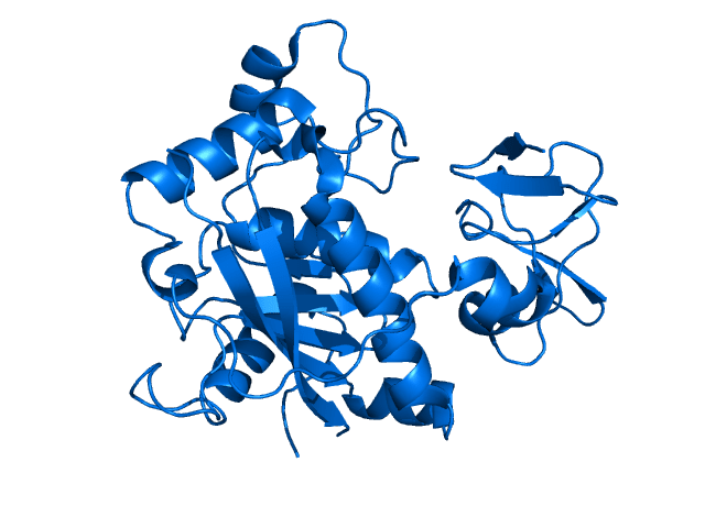
And, these findings pointed to ADAMTS4 as a molecular partner to DRFib activity. Single cell gene expression data had shown that Adamts4 expression was restricted to fibroblasts and endothelial cells before infection but was upregulated in fibroblasts exclusively after infection. This small enzyme is a matrix metalloprotease, meaning it degrades ECM proteins that build the scaffolding between cells. Specifically, ADAMTS4 degrades a loose extracellular network composed of versican called the provisional matrix, which is involved in cell infiltration and movement through tissue.
To understand what role ADAMTS4 plays in the balance of tolerance and resistance, the research team established a mouse knockout model for Adamts4 and challenged Adamts4−/− and Adamts4+/+ mice with a lethal dose of influenza virus. Mice without the enzyme had improved survival, and were able to maintain lung function at 6 days after infection. At 9 days, Adamts4−/− mice showed reduced alveolar inflammation and lung damage as compared to wild-type, again indicating a preservation of lung function. Comparing the CD8+ T-cell response to infection in knockout and wild-type mice, Adamts4−/− mice had a lower percentage of IFNγ-producing CD8+ T cells, though an equal overall percentage of influenza A virus–specific CD8+ T cells. Returning to a spatial view of infected lung tissue, the research team verified with immunofluorescence staining that there were fewer CD8+ T cells in lung sections of Adamts4−/− mice, especially in areas with intact versican, the glue of the provisional matrix and the substrate of ADAMTS4 (3).
Together, this indicated that knockout mice were still able to mount an effective virus-specific T-cell response, but with reduced inflammatory characteristics and immune infiltration in the lung and, thus, better lung function. And, ADAMTS4 was a crucial regulator of the T-cell response, in particular, the spatial localization of T cells in the lung, through modulation of the structural proteins that control T-cell movement in the tissue.
Understanding ADAMTS4 with real patient data
How does ADAMTS4 impact infection severity and outcome in human patients? To determine this, Dr. Thomas and his team took their observations to a clinical study of pediatric and adult influenza infection. Surveying the supernatant of endotracheal aspirate samples from 84 pediatric patients with severe flu, they noted that ADAMTS4 was the only factor other than age significantly correlated with disease outcome. In fact, levels of ADAMTS4 were all higher at the time of enrollment in the study for patients who had poor disease outcomes. In an adult cohort of 67 patients, they noted similar results, pointing to ADAMTS4 as a predictor of severe outcomes in influenza infection and providing further evidence for its role—degrading versican, and so opening the door to an overactivated, inflammatory T-cell migration into the lung (3).
A new therapeutic window for viral respiratory infection
How the body clears an infection is determinedly more complicated than the offensive activity of T cells and antibodies. As this study demonstrates, a detailed, functional view of the non-immune cells and tissue structures involved in regulating the immune response to viral infection is necessary to fully understand the factors that drive disease severity or, alternatively, preserve infected organ function and enable the best outcome for patients. Single cell and spatial technologies are helping scientists make these discoveries and pointing us, in the words of Dr. Thomas, towards a new therapeutic window for infectious disease:
“Limiting immune infiltration and maintaining ECM structure during influenza can improve the outcomes without compromising viral clearance. So we think there is a real therapeutic window here if it can be applied in a timely manner and in a rational manner to try to dampen some of these overexuberant responses to severely infected individuals'' (2).
Hear directly from Dr. Paul Thomas and explore the details of this study with our on-demand webinar. And find out more about the tools that supported this research with these resources:
References:
- Ayres JS and Schneider DS. Two ways to survive an infection: what resistance and tolerance can teach us about treatments for infectious diseases. Nat Rev Immunol. 8(11): 889–895, 2008.
- Thomas PG. Understanding Immune-Mediated Damage After Respiratory Infections. Webinar hosted by LabTools at The Scientist, and sponsored by 10x Genomics.
- Boyd DF, et al. Exuberant fibroblast activity compromises lung function via ADAMTS4. Nature 587(7834): 466–471, 2020.
