Optimal tissue section selection for spatial transcriptomics identifies crucial markers of mammalian embryonic development
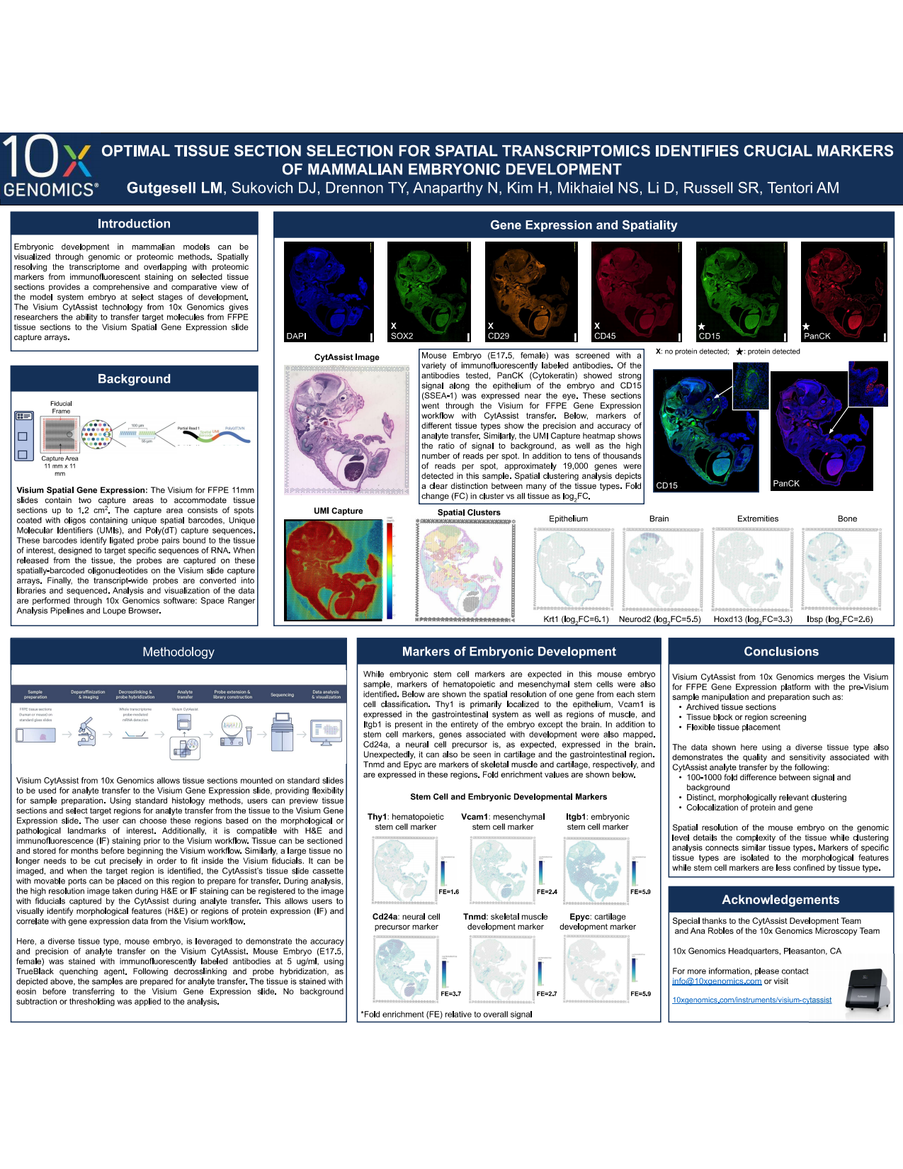
Embryonic development in mammalian models can be visualized through genomic or proteomic methods. Spatially resolving the transcriptome and overlapping with proteomic markers from immunofluorescent staining on selected tissue sections provides a comprehensive and comparative view of the model system embryo at select stages of development. The Visium CytAssist technology from 10x Genomics gives researchers the ability to transfer target molecules from FFPE tissue sections to the Visium Spatial Gene Expression slide capture arrays.
Embryonic development in mammalian models can be visualized through genomic or proteomic methods. Spatially resolving the transcriptome and overlapping with proteomic markers from immunofluorescent staining on selected tissue sections provides a comprehensive and comparative view of the model system embryo at select stages of development. The Visium CytAssist technology from 10x Genomics gives researchers the ability to transfer target molecules from FFPE tissue sections to the Visium Spatial Gene Expression slide capture arrays.