Why I chose spatial as a genomics core: Empowering spatial research for all
“We can take a project all the way through, and we won't turn you away because you have a different species or more challenging samples. We don't do anything easy.”
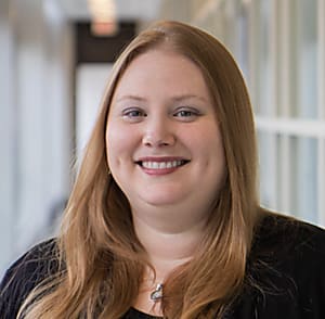
From skin to vein valves, the University of Michigan Advanced Genomics Core opens up its spatial services to even the toughest tissue types. In the words of its director, Olivia Koues, PhD, a former molecular epigeneticist and non-Hodgkin’s lymphoma researcher at Washington University (turned technophile for all things biology and sequencing), “Tiny tissue, large tissue, all organisms—we see everything.”

Recognized by the University of Michigan in 2022 with the Customer Service Award of the Year, the Genomics Core doesn’t just do hard things, but finds collaborative solutions and powerful technology to make hard things easier—and delight the researchers that make up their clientele with the highest quality data.
True to their roots, the lab offers next-generation sequencing services, bulk sequencing, and, since 2017, has supported single cell RNA-sequencing assays. “We now do over 1,500 single cell samples annually,” said Koues. In 2019, however, they made an unconventional shift to incorporate spatial transcriptomics assays, including Visium Spatial Gene Expression and Visium CytAssist, developing expertise in tissue sectioning and histology to make spatial experiments more accessible to their clientele.
We spoke with Dr. Koues to find out what it takes for a core lab to bring on spatial capabilities, the challenges her team had to overcome, and what they learned along the way. Keep reading to learn how spatial technology is empowering researchers to push the boundaries of their experiments and do more with precious samples.
What led you to bring on spatial assays at the Genomics Core?
It was a byproduct of our single cell success. The University of Michigan researchers are fantastic. They always want to push the limits and try something new. They were willing to jump off the cliff with us, in terms of bringing on spatial assays. And I was like, “Sure, why not, let's jump.”
What steps did you take to get started with your first spatial experiments?
We worked with Neil Winegarden, who was our Field Application Scientist at the time—Neil is fantastic. And we worked with various tissue processing facilities on campus to get the necessary components that we didn't have as a genomics core onboard. I met a lot of other core directors or facility directors, and they were extremely helpful.
What were some of the challenges you had to overcome to enable spatial assays?
Well, the pandemic hit. So we were able to turn out three projects before they sent us home and closed us down. And then they brought us back. I have 26 people that report to me in my facility, so we’re a really big core. I also had a lot of lab space. When they brought us back in after the pandemic, on shift schedules and with capacity restrictions, I was able to bring almost everyone in immediately, albeit on shifts. Some of the facilities we'd been working with for the upstream parts of the spatial experiments didn’t have that luxury. What we found was, when researchers wanted to come back and start doing Visium again—at that point it was a completely manual assay—we just couldn't coordinate between the two facilities. That’s when I got a cryostat.
From a genomics perspective—other than myself and maybe one other person—not many of us previously did tissue work. We had to hire experts in order to do that upstream experimentation. Originally, we thought that customers who are used to sectioning would section directly onto Visium slides and then pass them off, and we could take care of the rest of it. But that didn't happen often. Researchers were afraid to section onto expensive slides. It became a real challenge to get people through the onboarding process of what they were willing to do.
So that just led to a cryostat and a microtome. And as the assay evolved, we evolved with it. It was a matter of finding funds to procure instruments, or finding instruments that we could use that were not normally housed in genomics cores. I got an old microscope from the Microscopy Facility on campus that had all the imaging capabilities we needed for Visium. Then we upgraded it a little bit. We originally got a cryostat from a facility that had an extra one, and we've since upgraded that as well.
So we were procuring all this instrumentation that you wouldn't normally think would live in a genomics core, and then finding where to sit it. When you think of a genomics facility, you think a lot about pre-amplification, post-amplification—not so much sectioning and what room that needs to go in, right? What are the airflow parameters that need to be considered? Chasing little FFPE sections around a water bath is challenging if there's a heavy flow of air going.
We figured all that out. But what really helped was, about a year in, we hired a PhD-level histologist to come in. She spoke the antibody vocabulary, the tissue vocabulary, and the autofluorescence. So she's been fantastic to add to the core. We’ve since expanded and hired a pathologist and another histologist. We're growing that team, mostly because of the popularity of the assays. People keep asking constantly, “What can we do next?”
We had to develop the right skill sets, go outside the box, and procure other instrumentation that wouldn’t normally live in a genomics core. That's how we ended up becoming a Certified Service Provider—probably a little different than some of the other facilities around the country. We're doing the whole process—we’ll take blocks and section, and we’ll give researchers back processed data at the end. We're a full service facility.
How has the Visium CytAssist alleviated some of these workflow challenges?
We realized that spatial applications required more collaborative effort than our single cell projects. Single cells come to us as either live or dead cells, and we proceed or we don’t. But for spatial, there could be a lot of back and forth before the researcher actually pulled the trigger and said, “This is what I want to do.” We found that incorporating the CytAssist into the workflow actually streamlined that process.
Before we had a CytAssist, there were some challenges to taking on an entire project. A lot of external projects, and even some of the internal ones, don’t necessarily like passing off the blocks to us. Shipping blocks can be challenging, depending on the university or institute. We also spent a lot of time trying to explain to customers how they needed to intentionally embed their tissue, because we were going to section it onto a slide. If they wanted a particular part of tissue on the slide, they needed to be intentional about how they embedded it. When a block arrived, we checked the RNA quality, sectioned it onto a standard glass slide, then sent an image back to the customer and asked, “Was this what you expected to see?” Frequently, the answer was no. We had projects that took six months of back and forth on Zoom calls, and images going back and forth, trying to figure out exactly what they wanted to interrogate from a transcriptomic perspective.
The CytAssist has made this onboarding process a lot easier. For one, customers can now section onto the standard slide themselves—as long as they kind of hit the middle of a slide, they’re good. They're much more comfortable doing that. Shipping between places, or even around campus, is much easier. Also, once we do the staining and imaging of that tissue, if the researcher elects to proceed, we can go with that slide. We never have to worry about serial sectioning again.
How have spatial assays expanded the kinds of experiments you can offer to researchers?
Spatial applications have broadened the core’s projects to a whole new field. We got a lot of new customers that had never come to a genomics core before. They were straight up tissue people—pathologists, but also other researchers that had only looked at phenotypic changes in tissue histology images. We were able to bring them to the leading edge of science by incorporating genomics.
Now, people often want to do multiple assays from the same sample. Over the last couple of months, we've been actively working to combine Visium with single cell RNA-seq—doing it on serial sections, on scrolls. Being able to combine multiple assays and datasets has been empowering for people. Whether it's fresh frozen or FFPE—we've been using the Flex kit, too, with FFPE scrolls and have been happy so far with the data. It's allowing people to do more with precious samples.
We also have some customers that, for the Cytassist, have archived slides. Their tissue samples might have looked like junk to us when they started, but they proceeded because it was their one shot. This is the only slide they had in this one patient sample from however many years ago. Opening up to different sorts of samples that weren’t applicable for any of these spatial assays before has been a game changer.
One customer had been using a different platform, but they were waiting for the CytAssist. Because her tissue was so big—she just didn't have time to address how to cut it down to put it in a capture area and use it on the manual Visium assay. She was one of our beta testers for the CytAssist and has now switched everything over to the 11x11 mm squares on the CytAssist. We get a lot of patient samples from the operating room, like her samples. Being able to access old samples, but also the new ones, and to have multiple capture areas to configure based on your sample size have been nice additions to what we can offer customers to meet their needs.
How do the manual Visium assay and the CytAssist work together to enable more diverse spatial experiments?
Visium CytAssist is great for human analysis. With the manual Visium assay, there's some flexibility. We find the CytAssist workflow easier, but being able to do the manual assay with fresh frozen tissue—and it’s species agnostic—that's been really nice, because we have been able to accept samples that you just wouldn't be able to do with a probe-based approach at this time. We’ve seen some really nice canine data. We've done vein valves—those are really tiny. They only cover maybe 50 spots. Our customers are fantastic and they want to try anything. And it was successful—the data actually looked really cool at the end. Non-model organisms—we've been able to test them in difficult tissue types. We're not going to turn it away. We're going to do what we can to get customers data off their samples.
What are the most common projects and/or tissue types that you see coming in for spatial assays?
For the University of Michigan, we have a huge dermatology department. By far, our number one group for single cell and spatial happens to be dermatology. There's a big kidney consortium as well, so we do a lot of kidney. Prostate cancer and pancreatic tissue are also quite popular. We have a decent glioblastoma group on campus.
Again, spatial opened the door to investigators that didn't normally come to a genomics core, but a lot of our customers grew out of their single cell projects. A lot of them have huge single cell datasets that they can layer on top of the spatial assays that they are performing.
How do you educate potential customers on spatial assays?
Some researchers, like single cell users, are a little easier to onboard because they’re already familiar with tissue. They’ve been dissociating for a long time, and are also used to the data that comes back. Other users may have never approached a genomics core, so they don’t know what they might be getting back. They might understand the sectioning and the imaging part really well, and they might want to take on part of the workflow themselves, but the rest of it is a gray area for them.
So, we have a lot of consultations. That’s part of my job as the director of the core, to onboard new customers on the technology. We have 6 to 10 appointments per week where a customer will come to us with a project and they say, “I want to do spatial transcriptomics.” But they really don’t have any idea what platform or what assay to use. I spend time just going through the basics of the technology, how it works, what we would need from them, listening to their overall questions, learning about their biological systems, and helping to guide them down the right path forward. My job is to teach them about the technology and to make sure they’re picking the most suitable platform that will help them answer their questions.
We also do a lot of education and outreach—events, webinars. We found that data workshops are hugely popular here. So we’ll have our FAS walk through Loupe Browser data, and just get people the tools they need to start looking at their own data. They might still go to a bioinformatician at some point, but they’re the biological experts. So if we can teach them how to navigate the data, then we feel like we’re helping them down the right path.
What technologies are you excited to bring on next?
The request I get now is, “Better resolution for Visium.” We have a Xenium on order, so researchers are eager to incorporate Xenium. I think we can see, again, that synergy, the layering of multiple datasets. I get emails constantly: “Is it here yet, is it here yet?” And these are the same customers who would have been accessing us for either single cell or Visium at this point, so they’re just asking for the next thing that they can layer on their precious samples, and gather more data from.
This interview has been edited for length and clarity.
To learn more about the services provided by the University of Michigan Genomics Core Lab, please visit their website. And keep exploring resources for Visium Spatial Gene Expression, Visium CytAssist, and Xenium In Situ.
Want to find out why other researchers have chosen spatial? Keep reading the series.
