Single cell and spatial analysis of glioblastoma tumor organoids, a new tool for translation
Researchers from Washington University School of Medicine recently developed a brain organoid model of glioblastoma (GBM) tumors that recapitulates subtype-specific characteristics of patient tumors, introducing a validated human model to help expedite therapeutic development (1).
People with GBM are in desperate need of new therapeutics—even with treatment, the five-year survival rate is less than 5%. But extensive efforts to characterize the molecular drivers of GBM tumorigenesis and tumor progression have yielded few new therapeutics in the past decade.
Scientists have, however, made progress understanding the biology of GBM. Notably, extensive multidimensional analysis of over 600 GBM tumors by The Cancer Genome Atlas delineated clear subtypes of tumors, providing a framework for studying GBM biology and aiding diagnosis.
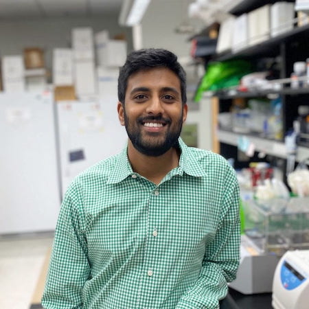
Matthew Ishahak, a postdoctoral fellow from Washington University School of Medicine, used this rich resource to engineer GBM brain organoids (eGBOs) generated from genetically modified stem cell harboring mutations commonly found in specific tumor subtypes.
We recently chatted with him about his preprint detailing the generation and validation of the newly developed eGBOs.

Read on to learn how Chromium Single Cell Gene Expression and Xenium In Situ were key in validating this new model system, and how Dr. Ishahak anticipates high resolution spatial transcriptomics technology like Visium HD will change our perspective of tumorigenesis.
What is the advantage to using eGBOs compared to other cancer models?
GBM tumors are super complex. Patients often don’t seek treatment until the cancer is very advanced, meaning their tumors have a high mutational load from the start. This makes studying the initial drivers hard to study with patient tumor samples, which are currently the most widely used model. Our eGBOs enable insights into early tumor initiation mechanisms. By introducing just a few mutations early on, we can see how that develops into these complex cancer phenotypes often found in patients. That’s what differentiates this model.
How does your model make it easier to study GBM tumorigenesis?
Glioblastoma tumors are classified into different subtypes based on certain characteristics, including specific sets of mutations. We generated two engineered stem cell lines using CRISPR to introduce specific mutations for two of these subtypes—proneural and mesenchymal. Both cell lines had a mutation in the telomerase reverse transcriptase (TET) promoter region, one of the earliest known mutations in GBM tumors. This mutation reactivates the NF-kappa B pathway, which isn’t normally active during brain development, and promotes rapid tumor progression. This allowed us to see the impact of the increasing loads of mutations that accumulate as the tumor progresses. Using these eGBOs, we can start to understand the effects of different mutations and the genetic drivers of intrapatient heterogeneity.
How did you determine your model was representative of patient GBMs?
We used single cell sequencing (Chromium Single Cell Gene Expression) and single cell spatial transcriptomics (Xenium In Situ) to see that eGBOs displayed expected differences in cell state and spatial architecture based on the initial mutation. First, we used single cell sequencing to validate that our eGBO cells had the same transcriptional profile as some of the cells observed in patients. Then, when we transplanted these organoids into mice, they formed tumors, which we didn’t see with normal brain organoids. When we visualized the tumors with Xenium, we saw our model recreated the spatial architecture of patients' tumors depending on which mutation was initially introduced into the stem cells. We were clearly able to see that different mutations impacted spatial organization differently. That was exciting to see because there isn’t a lot of spatial transcriptomics data available for GBM tumors.
If spatial transcriptomics isn’t widely used in GBM research, what led you to use it in your study?
Because my training is in biomedical engineering, I’ve always been interested in using new technologies. Traditional technologies—like immunohistochemistry (IHC) or PCR—require you to have an idea of what you are looking for ahead of time. You have to know what markers you want to stain for or what genes you want to amplify. Xenium allowed us to map expression of over 200 genes. We could find things that might be unexpected or that we didn’t know to look for ahead of time.
I also liked the ability to visualize gene expression across the entire sample rather than just protein expression of a small set of targets in limited areas as with IHC. We looked at how gene regulatory networks were dysregulated in eGBOs, not just individual genes or proteins. We were able to use all of this additional data to identify groups of genes that were co-expressed and determine what processes individual cells in different compartments of the tissue were really doing. This approach can help us identify pathways to target for future drug discovery studies.
Were there any results from your spatial analysis that surprised you?
With Xeniums’ subcellular resolution, we could see that cell interactions were altered in eGBOs. In wild-type organoids, proliferating cells and neuronal cells closely interact. But when we introduced oncogenic mutations, cell types from the tumor microenvironment—including endothelial and microglial cells—were interacting with proliferating cells instead. I don’t think we would have seen that without Xenium.
How do these altered cell–cell interactions impact gene regulatory networks?
That’s still an open question. It’s one of the reasons I want to continue this work with Visium HD. With Xenium, we were able to see cell types and gene expression of hundreds of genes. But to really delineate the impact of these interactions on gene regulatory networks, we need to be able to measure more genes expressed within those cell types. Visium HD will allow us to visualize the whole transcriptome so we can answer these open questions. These different cell types are spatially interacting, but what’s going on? We could use the whole transcriptome expression data to do things like inferred signaling analysis. We could not only look at what receptors are being expressed in one cell and what signaling molecules are expressed in a separate cell, but also look at if those two cells are close to each other spatially. We could infer if those signaling pathways are interacting with each other and link that to altered architecture. That would really help us intricately understand what’s going on in the tumor microenvironment.
How could this work impact drug discovery and development for GBM?
We wouldn’t have to rely solely on animal models or cell cultures that may lack the complexity we see in patients. If I start my own lab, I want to push in vitro models closer to replicating what we see in patients so that we can better characterize GBM with high resolution single cell and spatial transcriptomics. As we start to have a deeper understanding of the altered gene expression and spatial organization of these diseases using engineered models like eGBOs, that will provide another readout for drug screening. Having those additional redouts can only improve drug discovery. The more ways we have to tell if a target is working, the more chances it has to be successful throughout the whole pipeline, even into clinical trials.
This interview has been edited for length and clarity.
Learn more about this work in Dr. Ishahak’s recently published preprint. And find more resources about Xenium here and recently launched Visium HD here.
References:
- Ishahak M, et al. Modeling glioblastoma tumor progression via CRISPR-engineered brain organoids. bioRxiv (2024).
About the author:

