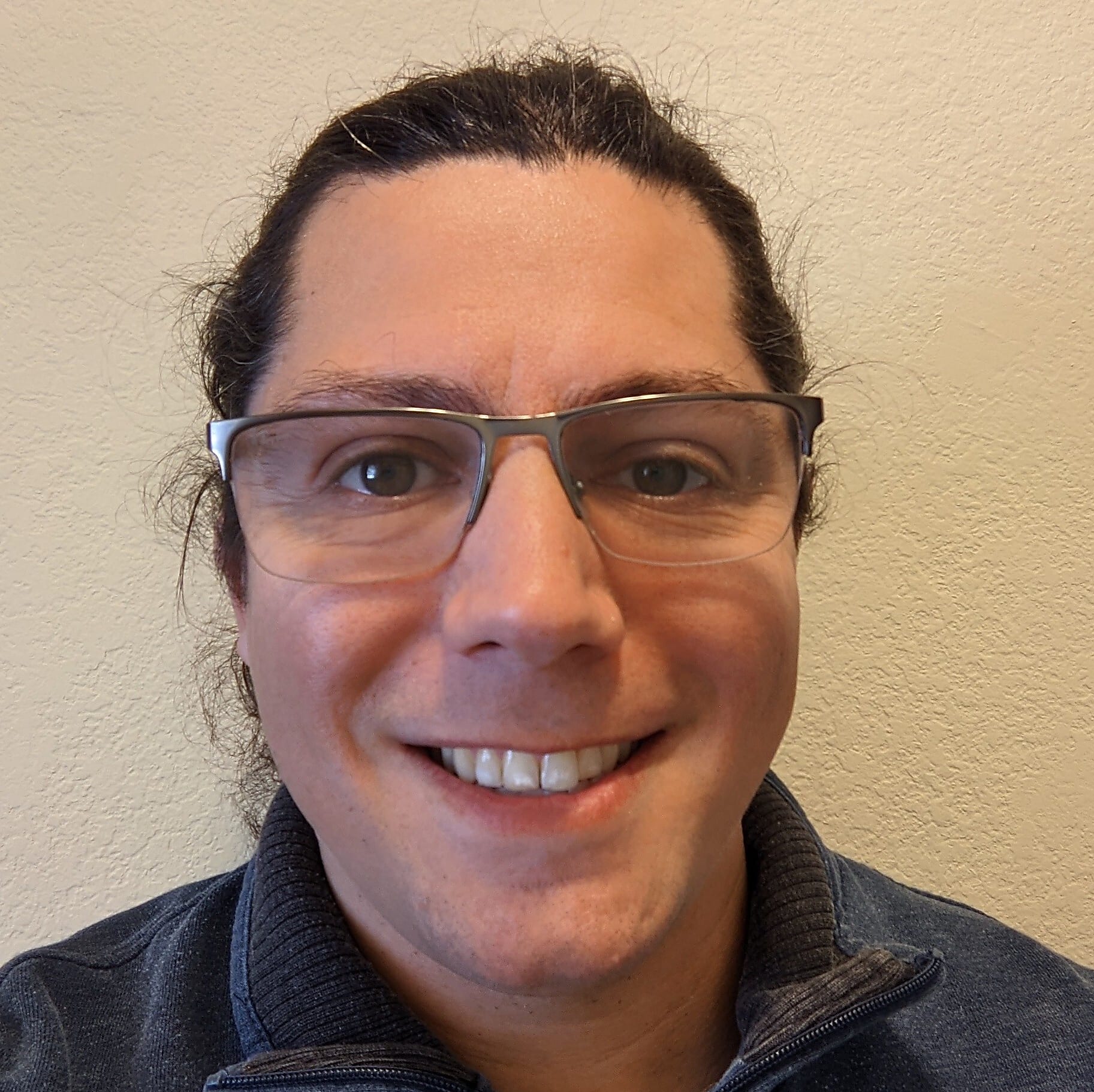
How Alon Millet ID’d the "what" and "where" of a new Alzheimer’s-associated cell type
Amyloid plaques have been recognized as a hallmark of Alzheimer’s disease for over a hundred years, but the mechanisms behind how amyloid may drive neurodegeneration remains quite a bit murkier. In an organ with as much anatomical, spatial, and cellular heterogeneity as the brain, an important question rises above all others: what cell types are involved in disease and response to amyloid?
Alon Millet, a graduate student researcher at Rockefeller University, had that same question and recently published his answer in Immunity. He was kind enough to share his insights on how:
- A novel microglial subtype may influence Alzheimer’s disease
- Bioinformatics in complex systems like neuroscience and immunology is evolving
- Single cell and spatial are making new and exciting approaches possible
*The Xenium platform is subsequently referred to as “Xenium” for the remainder of this piece.

Would you mind describing your research in your own words?
I'm an immunologist by training. My undergrad was all conventional, wet lab immunology with very little computation.
When I started grad school, though, I had this idea that a lot of the low-hanging fruit had already been picked. If you want to really discover new biology, especially in immunology, you really need to turn to at least some level of computation and high-throughput data collection to identify new emergent behavior in biological systems.
My work used computational biology as a jumping off point for two distinct questions. The first resulted in the Alzheimer’s paper, where I examined microglial heterogeneity in Alzheimer's disease across human and mice samples from organisms carrying the three different APOE isoforms. APOE is a critical gene that's a major risk factor for Alzheimer's disease; it’s very pleiotropic, it's immunomodulatory, and we don't really know why.
A major risk factor for Alzheimer’s disease is APOE4, and I identified a new microglial state that's strongly enriched in APOE4 carriers and becomes more enriched with age and disease progression. I showed that these microglia probably contribute to disease by being unable to clear amyloid beta and participating in inflammatory crosstalk with other immune cells. I also showed that currently existing therapeutics for Alzheimer's disease, such as aducanumab, clear this cell state back to a more functionalized role.
The second part of my work is still in progress. It uses single cell biology looking at physiological immunity in completely healthy mice and healthy individuals, and asks how common APOE mutants impact fitness and the state of the immune system. That work has led to some very interesting questions that are starting to turn mechanistic.
The common thread between these, though, was leveraging computational biology and using big single cell datasets to identify interesting phenotypes that can then be really explored at a more basic level.
How did you first become interested in researching Alzheimer's disease?
My laboratory actually studies cancer; we’re a metastasis lab. We are very interested in APOE as a factor that modulates a really wide range of biological functions. For instance, at the peak of the pandemic we had a paper showing that APOE variants impact COVID mortality.
My perspective coming into the lab was that we really don't understand how APOE does these things. We see APOE pop up in all these different systems and disease states, but we have no clue what it’s up to. So I thought, where better to look than a really well-defined system where we know APOE has these big impacts? So even though we're not an Alzheimer's lab, we thought this was a good place to look to better understand APOE.
How do you think that the single cell and spatial tools you used were helpful in addressing the shortcomings of Alzheimer’s disease models?
Both single cell and spatial were essential to the project. It would not have been feasible with conventional tools. Single cell is essential because we're studying a population that's incredibly rare: this microglial state only comprises 60–70% of microglia in 2 year old mice and only about 0.1% of microglia in young mice, and microglia themselves are only about 1% of cells in the brain. It would have been impossible to identify the cell state without single cell techniques.
Once we found that cell state in mice, we had to try and identify it in humans. We originally thought we’d do single cell again but, to make a very convincing claim that these cells are real and to understand what they're doing in the tissue, we turned to spatial since it’s a very powerful technology for that.
We took human Alzheimer’s tissue samples and asked whether we saw those cells in human samples—which, fortunately, we did. A lot of Alzheimer’s disease models don’t recapitulate the human disease, so it was a big relief to see those cells in humans. That let us ask really critical questions: where are those cells in the brain, what is their local niche, and what cells are they surrounded by?
After running these tissues on the Xenium platform, we were able to stain the same sections for amyloid beta given that Xenium is non-destructive. We saw the local environment around this microglial subtype was enriched for amyloid beta. This tipped us off to a potential mechanism, and we followed up to confirm that they’re no longer capable of clearing amyloid beta as efficiently as normal microglia.
The ability to really look at the local niche around these cells in situ and undisturbed—to not have to do digestion, which can really stress microglia out—was a really, really critical component of the study. It made a lot of our discoveries feasible in the first place.
What led to your decision to apply single cell multiomics and Xenium single cell spatial imaging to Alzheimer's disease?
We used three big tools over the course of the paper. The first was single cell sequencing with Chromium Universal 3’ Gene Expression to see if there were changes in heterogeneity in APOE2, -3, and -4 microglia over the course of Alzheimer's disease progression.
The next tool that we used was multiome single cell sequencing with Epi Multiome ATAC + Gene Expression to identify not just the transcriptional state and how it varies in this model, but also what happens to the epigenome. This approach was critical for us to identify molecular features and drivers that produce this state in the first place.
Having both single cell ATAC- and RNA-seq data from the same cells let us build some really nice computational models of the gene regulatory networks that undergird each of these different microglial states, and that lets us identify putative transcription factors that influence their state.
Spatial was a natural extension of that. We’d found the cell state, now we wanted to know what it’s up to, what it’s surrounded by, and use that to figure out phenotypic outputs. Spatial tipped us off to the fact there are differences in their surroundings with respect to amyloid beta.
We were able to look at the niche around these cells and find that they're surrounded by astrocytes that overexpress amyloid precursor protein (APP). So you see that one of these stressed microglia is literally always right next door to one of these astrocytes that overexpress APP.
This kind of analysis is only possible through spatial, and it led us naturally to these next steps of a potential disease mechanism.
Researchers tend to not be bioinformatics experts, particularly in pathology and clinical neuroscience where there’s a lot more use of legacy methods. How big of a barrier do you think bioinformatics is for researchers trying to answer these next-level questions?
I think this speaks to the bigger image of how bioinformatics has shifted over the last 30 years. Back then, there were wet labs and there were dry labs, and they didn't really mix at all. If you go back 15 years, you would have collaborations between wet and dry labs, but, still, they were kept separate. If you go back 5 years, you start to see both people in a single lab. What we’re seeing now is a single person is expected to do both the experiments and the analysis afterwards.
I think tools like Chromium and Xenium are a natural extension of that because they democratize a lot of these technologies that would be prohibitively expensive and difficult if they weren’t wrapped nicely into packages. Similarly, there’s so many tools out there from exclusively computational biologists who’ve developed technologies and tools that let you easily analyze this sort of data at these scales.
What were the biggest challenges you had to overcome in your research approach?
Microglia are very sensitive so, depending on how you digest your tissue, you can completely skew the transcriptional profiles that you get out. So that took some optimization.
I think the single trickiest experiment that we did was from Figure 6 of our paper, which was the follow-up to our Xenium experiment. The Xenium data showed us that the immediate neighborhood around these microglia is enriched for amyloid beta, suggesting they’re deficient at clearing amyloid compared to other microglia.
We wanted to know: if we take these microglia out of the brain and incubate them with amyloid, will they take up more or less amyloid? We needed a true functional experiment, and it turned out to be very hard because we had to optimize a lot of things about the system. So it was technically demanding, but it was worth it because it ended up being a key feature of the story that we have this clear functional demonstration that these microglia are impaired.
What do you think the next steps are for your research, and what would you like to do next?
Following up on the Alzheimer's story, there are two questions I’m really interested in. The first is to what extent the microglial subtype I identified is also relevant to non-Alzheimer’s disease neuroinflammation, such as multiple sclerosis, brain tumors, etc.
Question number two is whether, and to what extent, these cells are a therapeutic target. If we had some way to reliably revert these cells back to a more functionalized state, could that be therapeutic?
Finally, we’re interested in how targeting APOE might impact the emergence of these cells. There’s a company associated with our lab, and one of their products is an APOE agonist. We’re curious about what happens if we give this drug to Alzheimer’s model mice, to see if it has any impact on disease progression.
I also alluded to my interest in clinical trials and translational findings. I think spatial offers the opportunity to do more and better drug testing, better drug screening—getting a better idea of what’s going on in the brain.
What do you think are the most promising future applications for spatial transcriptomics and neurodegenerative disease, especially in areas like pharma?
When it comes to pharma, you're looking for scalability. You have a huge number of potential drugs that you're trying to screen, at least early in the process. Conventional single cell is probably the best tool at this stage because you have more scalability.
I think, once you have a smaller number of drug candidates, that's where spatial can be powerful. You can go into a system of interest and say: I have 5 or 10 promising drug candidates that I'm curious about, but now I want to understand them in the context of the organism. You can do it either by having a large tissue section or, if you want scalability, as a tissue microarray.
You can tile your slide with sections from a bunch of different samples to have biological replicates, or treat with a bunch of different drugs all together on the same slide and get a huge amount of data very cheaply. There’s real power there.
For instance, when I was doing my single cell analysis, I was getting maybe 15–20,000 cells total from a single chip. With Xenium, however, a single slide gave me almost 200,000 cells because it's imaging based, so scalability from a single section is much higher.
It lets you answer questions with a lot more biological and statistical power. You can ask questions that aren't possible to ask without these tools: not just the effect a drug has on a cell type of interest, but the effect it has on all the cell types across the tissue. You can ask about the location of the cells that were strongly impacted by a drug, what happens to the cells around it, how it changes cell–cell communication. When you have spatial data, that becomes much easier because you can see which cell is next to which and better model the disease network.
Is there anything really cool or interesting you’d like to talk about that you haven’t gotten a chance to mention yet?
Something that I’ll be doing very soon, which I think is enormously powerful, is Perturb-seq. I think it’s a method that’s only recently started to pop up on people's radars, where you can do single cell sequencing in combination with CRISPR screening.
5 or 10 years ago, you’d have a pool of cells that you’d transduced with CRISPR guide RNAs, then you’d sort them to get a binary signal: cells either responded, or they didn’t. Then you’d sequence your guide RNAs and see where you had enrichment.
But we know biology isn’t binary so, when you structure your output like this, you mask a lot of your ability to understand what’s going on in the system. So, with Perturb-seq, your output isn’t binary, it’s the whole transcriptome because you do single cell sequencing plus guide capture. It’s incredibly scalable and it gives you so much information from a single experiment on exactly what's driving your phenotype at a level that wasn't possible before.
With how cheap sequencing is getting and how much better these tools are getting, you have the birth of this new paradigm in biology where a single experiment can almost entirely answer a biological question on its own. Especially now that people are combining this with multiomics where you can do single cell screening, capture your guide, capture your RNA, capture chromatin modifications, and capture surface protein levels. You get a full picture of everything in your biological system right out of the gate. This wasn’t possible before.
So I'm excited about that space, and there are some much smarter people than me designing some really cool experiments using these technologies to answer questions that you simply couldn’t before.
“What” and “where” lead to a how: How single cell and spatial technologies are reshaping neurodegeneration
In this interview, Alon Millet highlighted how he used multiomic single cell sequencing to identify a population of terminally inflammatory microglia in an Alzheimer’s disease mouse model, then both validated their existence and localized them in human patient brains.
His approach to unify both the cellular and transcriptomic “what” and the spatial “where” is critical for understanding the complexity of the brain and represents the latest chapter in a history of new technologies leading to Alzheimer’s advances. We’ve known about Alzheimer’s disease for over 100 years, but we’ve only had PCR for 40 years, NGS for 25 years, and single cell sequencing for 15 years. Now, we have both imaging- and sequencing-based spatial transcriptomics.
Imagine what more we’ll discover in the coming years with approaches similar to Alon Millet’s.
To discover more about the technologies used in this paper, check out Epi Multiome ATAC + Gene Expression and the Xenium In Situ platform, or see what more Xenium single cell spatial imaging can do in Alzheimer’s disease models in our Application Note.
Questions? Contact sales.
