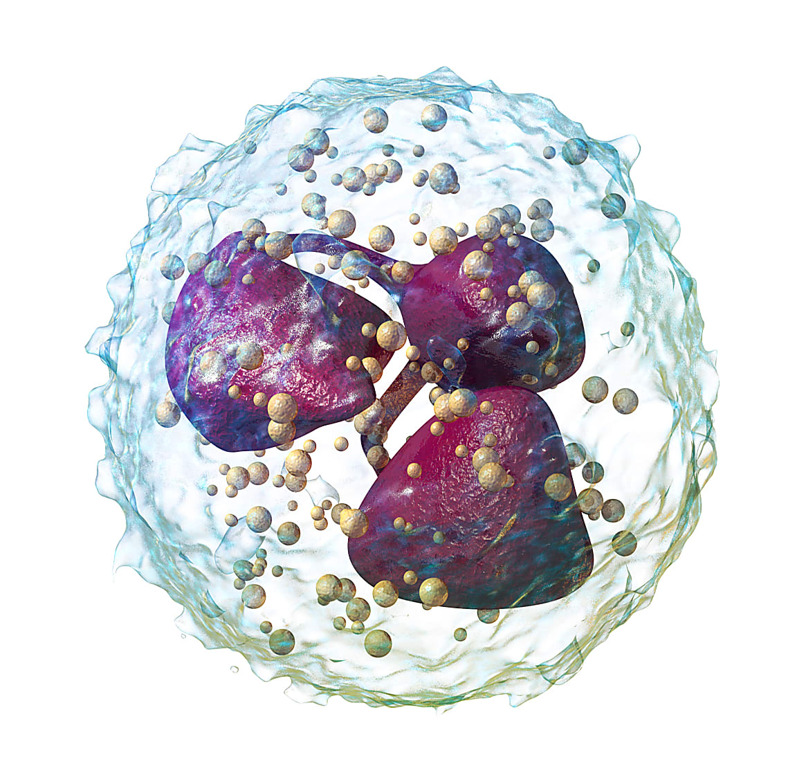
Tracking neutrophil development along a continuous spectrum with single cell analysis
While neutrophils are one of the most common types of white blood cell and perform an essential function in innate immunity—phagocytosing pathogens—they remain somewhat of a mystery. To a large degree, these cells vary when it comes to many different factors, including morphology, surface markers, and migratory, phagocytic, and suppressor functions (1). This makes them hard to classify. Importantly, characterizing subpopulations of these cells is difficult because, within a heterogeneous mixture of neutrophils, there exist cells that appear phenotypically unique based on differentiation state. In a recent paper (2) published in Nature Communications, scientists led by Ricardo Grieshaber-Bouyer, MD, at Harvard Medical School and the ImmGen Consortium (Immunological Genome Project) set out to discover more about the nature of neutrophil differentiation and activation using single cell transcriptomic profiling.

Neutrophils are some of the most abundant cells of the innate immune system, making up anywhere from 40 to 70% of all white blood cells in humans. While mature neutrophils reside in the blood, they are derived from bone marrow and can differentiate into various short-lived subtypes. In this work, Dr. Grieshaber-Bouyer and team used a combination of single cell RNA-seq (scRNA-seq) and computational analyses to characterize a total of 17,000 mouse neutrophils, finding that within populations, cells exist not in discrete subtypes but along a phenotypic continuum depending on their stage of differentiation. They call this continuum “neutrotime.”
Neutrophils exist along a continuous spectrum of developmental stage
To dig deeper into how neutrophils exist among a mixture of cells, the team performed scRNA-seq on 12,672 healthy mouse blood, bone marrow, and spleen cells. Clustering analysis allowed them to classify four subpopulations based on development stage (P1 was the earliest, P4 was the most mature), and that the subpopulations were distributed differently across tissues: P1–P3 were most abundant in the bone marrow and P4, in the blood and spleen. Using Monocle analysis, which is built around the concept of pseudotime, or, a “measure of how far a cell has moved through its developmental or biological process,” they revealed a continuous distribution of neutrophils across bone marrow, blood, and spleen. Because this analysis did not include pre-neutrophils, they took a diffusion map approach for further analysis.
Applying diffusion maps, a “dimensional reduction method that orders cells based on transition probabilities and is sensitive to branches in the data (3–6),” they found that neutrophils across all tissues could be mapped onto a spectrum. Cells from bone marrow resided at one end of this continuum while neutrophils in blood and spleen were at the opposite end. Bone marrow contained mostly immature cells; spleen contained neutrophils at all stages, but had an abundance of later-stage but still somewhat immature neutrophils; and in the blood, the majority of cells were mature. RNA velocity vectors uncovered a unidirectional progression from one end of the continuum to the other. (RNA velocity is a method that “compares unspliced and spliced mRNA to provide an unambiguous time vector to transcriptomic data (7).”)
Transcriptomic profiling reveals clues to neutrophil activation
Their scRNA-seq data showed that gene expression differences between states of neutrotime are continuous, without discrete breaks. In other words, some genes increase in expression with neutrotime while others decrease. Gene Ontology analysis showed early neutrotime genes were associated with anabolic functions, including metabolic processes, and defense response. Late neutrotime genes were enriched for cellular responses to the environment, including immune responses, the cellular response to toxic substances, and gas transport. Comparing Human Cell Atlas data, they saw that human neutrophils show a gene expression pattern mostly consistent with neutrotime in mice.
They also saw changes in the type 1 interferon response in neutrophils, depending where the cells were along the neutrotime continuum. Ifitm2 and Ifitm1 increased with neutrotime, Ifitm3 was expressed throughout neutrotime, and Ifitm6 was expressed early in neutrotime. By contrast, genes associated with type II interferon response were expressed in low amounts and showed no pattern.
The gene expression changes seen during the progression along neutrotime reflected the change in transcription factors (TFs) over the course of development as well, with different TFs active in early versus late neutrotime. Specifically, lactoferrin (Ltf, both a TF and a secondary granule protein) and Cepbe were strongly active in early neutrotime. By contrast, later neutrotime was characterized by the activity of TFs encoded by Atf3, Klf2, Cepbb, Junb, and Jund.
Inflammation leads to polarization and gene expression changes in neutrophils
To understand how gene expression differs across neutrotime during inflammatory states, they performed scRNA-seq analysis on several types of mouse neutrophils, K/BxN serum–induced arthritis (blood and joint); IL-1β pneumonitis (lung); and IL-1β peritonitis (peritoneum). Analysis showed that cells from inflamed tissues polarized either toward acute IL-1β–induced inflammation (lung or peritoneum) or sub-acute K/BxN arthritis. Arthritis blood neutrophils were less mature than healthy blood neutrophils, suggesting early release of neutrophils during tissue inflammation. Neutrophils from inflamed lung and peritoneum were immature, suggesting that less mature neutrophils are recruited to acutely inflamed tissues. Interestingly, IL-1β–inflamed lung and peritoneum differed from each other in 224 transcripts, pointing to gene expression varying with site of recruitment. They also identified different TFs associated with different kinds of acute inflammation, highlighting the changing nature of gene regulation in mature neutrophils as they emerge from neutrotime.
A new framework for understanding neutrophil activity
This intriguing blend of single cell and computational analyses provides valuable insight into heterogeneous populations of neutrophils, specifically that, it’s not different kinds of neutrophils that exist, but different states of maturation and activation of a single cell type. As a working model, they believe that as neutrophils meet environmental perturbations, their gene expression patterns change based on cell and tissue location, type of stimulus, and time, leading them to take on a specific phenotype. This new framework will be useful for scientists aiming to further understand neutrophil activity during both health and inflammation or disease.
For more information or to access their data, visit immgen.org.
References
- Ng LG, et al. Heterogeneity of neutrophils. Nat Rev Immunol 19: 255–265, 2019.
- Grieshaber-Bouyer R, et al. The neutrotime transcriptional signature defines a single continuum of neutrophils across biological compartments. Nat Commun 12(1): 2856, 2021.
- Angerer P, et al. Destiny: diffusion maps for large-scale single-cell data in R. Bioinformatics 32: 1241–1243, 2016.
- Haghverdi L, et al. Diffusion maps for high-dimensional single-cell analysis of differentiation data. Bioinformatics 31: 2989–2998, 2015.
- Moignard V, et al. Decoding the regulatory network of early blood development from single-cell gene expression measurements. Nat Biotechnol 33: 269–276, 2015.
- Haghverdi L, et al. Diffusion pseudotime robustly reconstructs lineage branching. Nat Methods 13: 845–848, 2016.
- La Manno G, et al. RNA velocity of single cells. Nature 560: 494–498, 2018.
