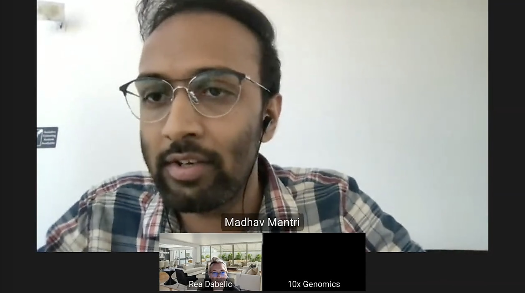
5 expert tips for difficult single cell tissue dissociation: A conversation with Madhav Mantri
Single cell tissue dissociation is not necessarily a one-size-fits-all process. Depending on the tissue type, species, sample age, processing environment, and other factors, tissue dissociation for single cell experiments can be difficult, requiring practice and optimization to produce the best results. But the possibilities of discovery are well worth the effort, and there’s help along the way.
We featured Madhav Mantri, PhD candidate from Cornell University, at our recent Immunology Spotlight Webinar where he discussed his personal experiences dissociating mouse heart and ileum samples for a Chromium Single Cell Gene Expression experiment. As someone who has already gone through the process and found what works for his samples, he offered a number of valuable tips for scientists interested in performing their own single cell experiments. Read on to find expert guidance for difficult single cell tissue dissociation, then watch the on-demand webinar or explore methodological details in the research team’s pre-print to learn more.
Tip #1: Practice, practice, practice
How to practice well and achieve success during the real experiment—when precious samples and reagents are truly on the line—were Mantri’s first comments in his conversation with 10x Genomics Sr. Market Development Manager for Immunology, Rea Dabelic, PhD, at our Immunology Spotlight Webinar.

Mantri’s research focuses on viral-induced myocarditis, inflammation of the heart caused by infection (check out the blog summary here). Leveraging a neonatal mouse model of Reovirus-Type1-lang infection, he resected heart and intestinal ileum tissues from mouse pups across multiple time points over the course of infection.
“I had to do a lot of practice first, with mock mice, not the viral-infected mice I was going to use, because I didn’t want to put all that work into infecting these neonatal pups with the oral gavage mechanism and then not use that tissue for the experiment. So I practiced with mock pups of the same age, almost having the same tissue size.”
Beyond practicing with age- and tissue-matched samples from model organisms, Mantri also recommended how to find comparable practice tissues when working with precious human samples. This is a catch-22 of biological research: while it is essential to practice before using precious human samples, due to the limited sample amount you’ll have, it is difficult, if not impossible, to actually find human practice tissues (because they’re precious).
Mantri suggested starting with scientific literature to first define the extracellular matrix (ECM) components in the tissue you want to work with at the age of the donors or patients you’ll have access to, as that will heavily influence the tissue dissociation protocol. From there, he recommended practicing with an age-appropriate mouse tissue: “Find in what aged mice you would have the same ECM components, and use that tissue to practice dissociation.”
Dabelic added to these suggestions, pointing out that the Human Cell Atlas consortium provides a data portal cataloging a number of different human tissues, including protocols that were used for single cell tissue dissociation. She noted that this could be a valuable tool for reviewing those protocols or getting into contact with the scientists who performed them, as they are experts at working with those tissue types for single cell experiments.
Additionally, Mantri offered a tool that he uses often in preparation for experiments—the 10x Genomics publication page, a catalog of peer-reviewed publications using 10x Genomics technology, with filtering capabilities according to tissue type, species, and other fields. This provides a menu of publications to review when searching for methodologies that match the samples you want to work with. (And, of course, you can always reach out to 10x Genomics support for help, too!)
Tip #2: Use best practices for tissue collection and processing
Mantri walked us through his experiment, from tissue collection to Eppendorf tube, making note of best practices for sample processing. First, because he was using the fresh-frozen single cell RNA-seq kit, he planned his experiments early and was sensitive to the environmental needs of the fresh tissue to optimize cell viability. Focusing on heart tissues, he said:
“I would get the heart tissue, and then put it in the plate with ice cold HBSS solution. Then I would perfuse the heart and make sure I remove as much blood as possible from the chambers. Then I would transfer the tissue to a new plate, where I would mince it into tiny 1 mm square pieces. And during all of this, the tissue has to be maintained at ice cold temperatures, in ice cold wash buffers, and should not go to room temperature at any time.”
Mantri shared additional details: “I did it all on plastic* petri dishes, which were lying on huge ice buckets.”
*10x Genomics now recommends the use of uncoated clean glass dishes instead of plastic for tissue mincing to minimize the presence of solid debris in cell suspensions. For more details please review the following document.
(We’d like to pause here to appreciate both the effort required to do this, and the visual image it’s bringing to mind. Claps to you, sir. Continuing…)
“I washed the heart tissue in one plate, and then I used forceps to lift it and put it in a fresh solution. Now I know the washing part is done, so I have to start mincing. I would remove most of the HBSS to get the tissue semi-dry. I would not submerge it in a lot of liquid, but have a little bit of liquid solution HBSS in the corner of the dish, put my tissue there, and then use the blade right there to chop it on the petri dish. Then I would use a transfer pipette to take the tissue in, with the solution, and transfer it to an Eppendorf tube. That’s why I would not use a lot of solution [in the petri dish], because I use the tabletop centrifuge to wash the minced pieces before I start the enzymatic dissociation protocol.”
Practice is also essential for sample collection and processing, as Mantri emphasized, because some tissues are more difficult than others to remove while maintaining good tissue quality. For example, intestinal ileum tissues begin to digest as soon as the animals are sacrificed, making it necessary to work quickly to get them into ice cold solution. In some cases, when the tissues are hardier, such as with the heart, it can be easier to perfuse them while still in the animal.
Tip #3: Tailor enzyme cocktails and reaction times to cell type and tissue
Enzymatic dissociation is the next key step to release single cells from minced tissues. According to Mantri, there is a “sweet spot” when it comes to enzymatic tissue dissociation: “You cannot take too long to dissociate the tissue because that is going to affect your cell viability. But then you cannot do it too fast by adding too much enzyme because the enzyme reaction also happens in conditions that are not favorable for the tissue.”
A time series experiment can help to determine the right amount of enzyme to use over a certain timed reaction:
“For lower times, you use higher concentrations of enzymes; and for higher times, you use lower concentrations of enzymes. You can make a table with enzyme concentrations and time courses. Split the tissue into different tubes and do these reactions, then do cell counting at the end to find out your viability percentage. I did that in the beginning for heart tissue, so I knew I needed to do at least a 30-minute dissociation.”
Mantri emphasized that finding the sweet spot for dissociation requires special attention to the cellular composition of the tissues.
“Dissociation is a cell type–dependent process. Different cell types have different levels of tolerance to stress. For example, myocytes are cardiac-specific cell types that have to be handled very carefully, otherwise you won’t get a very good yield...Optimizing the dissociation for myocytes and smooth muscle cells is very important because those die very quickly.”
There are a number of available resources to guide you as you make this determination on the right enzymes and concentrations for your specific samples and the cells they contain. Mantri used a database provided by Worthington Biochemical Corporation, offering cardiac tissue–specific recommendations for enzymatic dissociation, which he highly recommends to others. Additionally, Mantri suggested that if you’re looking to study a specific cell type from the sample, such as stem cells, it can be efficient to leverage commercially available kits to isolate that cell type of interest.
Dissociation can, however, become more complicated when the same tissues have conflicting enzymatic needs. For example, Mantri found that ileum tissues were more complicated than initially expected: “the gut is made of completely different cell types than the villi,” which influences the efficiency of the enzymatic reaction. Additionally, the gut tissue itself is intolerant to some components of the basic reagents Mantri was using for other tissue types in his experiment, PBS and HBSS. “The dissociation enzymes that we use to break the fibroblast matrix [in the gut] should not have [Trypsin-EDTA]. But it is used to get the villi off the gut, very quickly.” In this case, EDTA was a double-edged sword: it worked perfectly to dissociate the villi, but inhibited the activity of collagenase, the enzyme Mantri wanted to use to dissociate the gut wall tissue, by neutralizing calcium and magnesium ions in the solution which were necessary for collagenase to work.
Mantri described a workaround that involved dissociating the gut and villi separately:
“It was taking more than an hour to get the wall tissue dissociated. But when I did it in two steps, and got the EDTA and the villi epithelium cells out—I kept them on ice and took the chunks of the wall tissue that were left over and transferred them to a new tube where I washed them thoroughly to get the EDTA off. Then I put in the collagenase and supplemented it with calcium chloride—then I found out it can be done in 20 minutes.”
These innovative modifications are the result of trial and error, even some failed runs for Mantri. However, the lessons are now invaluable for his and others’ future experiments. “Sometimes dissociating the same tissue could have conflicting reagent requirements, so you have to be careful that you wash the stuff that is limiting the enzymatic reaction in any way before you start the next reaction.”
Tip #4: Consider using serial enzymatic dissociations
Another pro-tip that Mantri offered to both accelerate the dissociation process and ensure optimal cell viability is a method called serial dissociation. “Doing a serial dissociation is a very effective way to speed dissociation up, but not by increasing the enzymatic concentration.” Mantri explained that he starts with the minced tissues in a 2 mL Eppendorf tube mixing on a thermocycler at very low speed. He described the process in further detail:
“After every 10 minutes in a 30-minute dissociation protocol, so 3 times during the protocol, I would spin the tube down or let it settle, removing it from the thermocycler and keeping it stable on the bench. I let the big chunks of tissue settle down, take the suspension off, and put it in a new tube on ice. If there are some cells out of the tissue, in the solution, I don’t want them to stay unhappy for 30 or 40 minutes [during the rest of the dissociation protocol]. I want them to go to a happy place on 4 ℃. So I would have a bigger tube ready with a lot of happy solution, basically a lot of PBS + BSA, which you have to eventually resuspend the cells in. Then, I would replace the enzymes with fresh enzymes to speed up the entire protocol. If you’re using the same enzymes for 30 minutes, the activity of the enzyme is not the same during the dissociation protocol. If every 10 minutes, 3 times, I change the enzyme, that speeds up the dissociation in the next round. The dissociation would happen more efficiently and you’ve already taken out the cells that are in the suspension form and put them in a happy solution, PBS BSA.”
Tip #5: Remove red blood cells earlier rather than later
After single cell suspensions are formed, there are still a couple of crucial housekeeping steps before loading cells into a Chromium instrument. It’s important to wash the cell suspensions to remove unwanted cells or debris, particularly any remnant red blood cells from the tissue. Mantri shared what he does to ensure clean cell suspensions:
“I typically use red blood cell lysis buffer (ACK lysing buffer) to remove the red blood cells. One important thing I would say there is, if you’re doing 3 or 4 wash steps, do the red blood cell lysis sooner rather than later. As red blood cells are lysing, they release their RNA into the solution. If you are not in 50 mL tubes at this point, but 15 mL tubes, it makes a big difference if you wash out any free floating RNA from the solution. If you don’t wash it off enough, and there is free floating RNA in the solution, and you load that into the Chromium instrument, those RNA molecules will get barcoded in the droplets and you’ll have background everywhere in your data.”
Mantri emphasized that it’s essential to be diligent about washing steps even before tissue dissociation begins, to ensure that as many unwanted cells are removed at those stages, just as they will be at this step of red blood cell lysis.
We’d like to give a big thank you to our speaker Madhav Mantri for sharing these helpful insights, straight from his own experience of running single cell experiments. If you want to hear more, watch the Immunology Spotlight on-demand webinar here. There’s always more that can be said about how to optimize these protocols, so please explore our User Guides and support documentation to get more detailed information and guidance.
