Now shipping
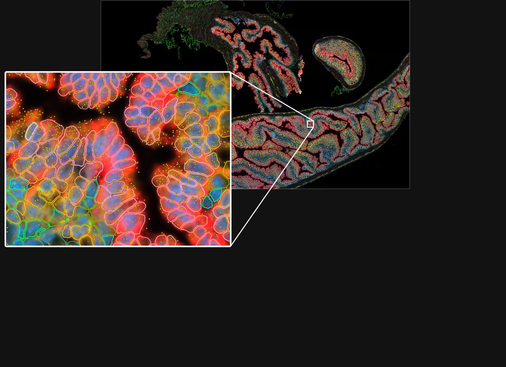
A new standard for cell segmentation
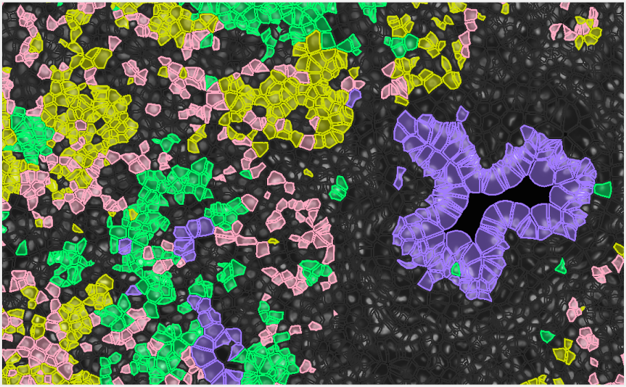
- Have more confidence in transcript-to-cell assignments with precise cell segmentation using multiple morphology features
- Integrate easily into your Xenium workflow using a simple stain mix
- Reflect cell morphologies more accurately with the advanced AI algorithm
Built for rapid data exploration
The Xenium Analyzer processes data during the run allowing you to visualize and explore the data right away.
Use our open file formats with community-developed tools of your choosing.
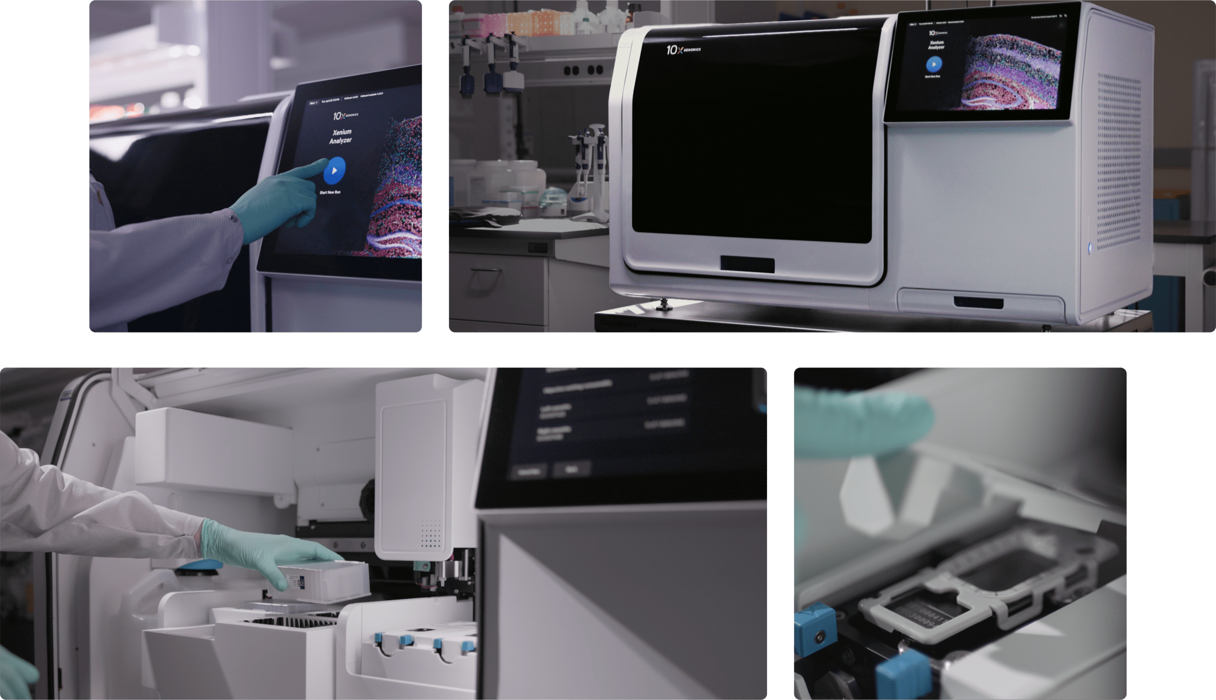
A state-of-the-art single cell spatial imaging platform
Xenium Analyzer integrates high-resolution imaging and onboard data analysis enabling the processing of 400 mm² of tissue in <50 hrs (with nuclei-based segmentation).
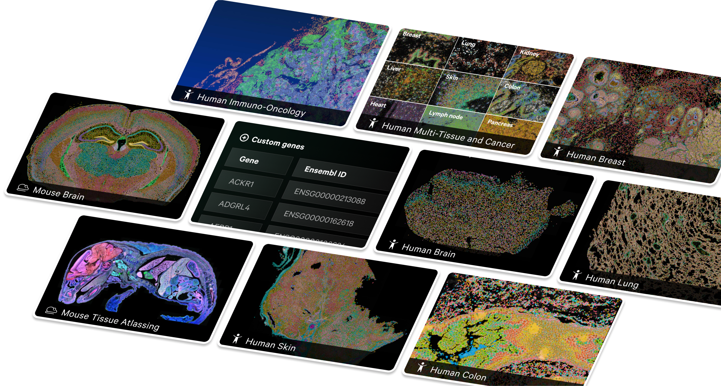
Target the biology that matters
Our panels are carefully designed and curated, incorporating a data-driven approach that combines expert input with years of single cell experience. Customization is fast and easy.
Resources
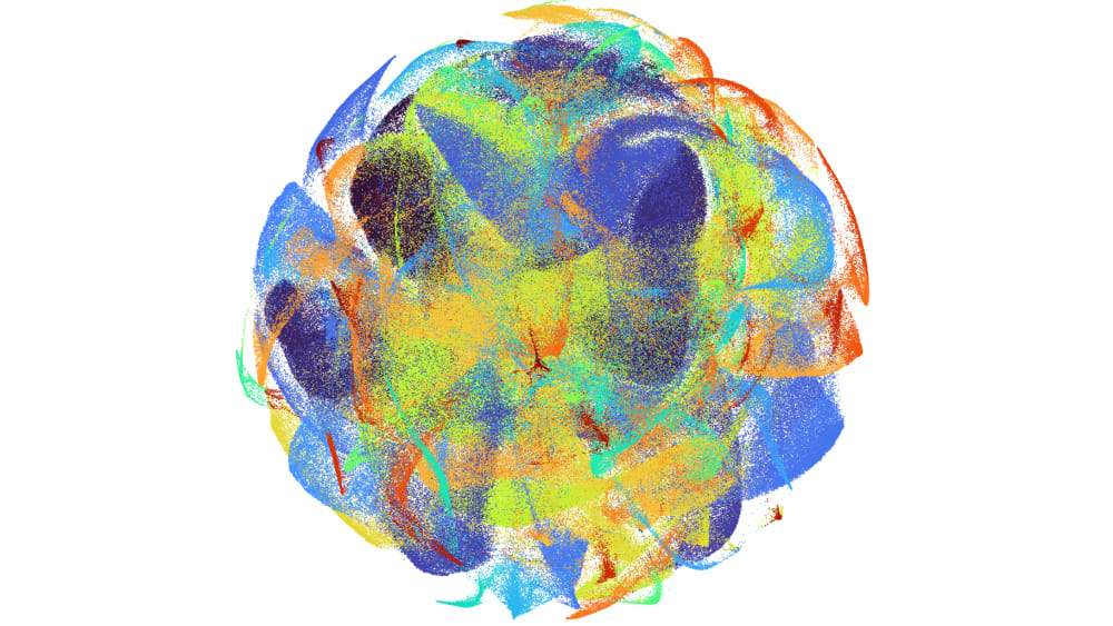
See Xenium data in action
Explore our 474 gene human pancreatic cancer dataset post-Xenium H&E and IF images.
Grant writing assistance
Get the information you need to write grants featuring Xenium experiments.
Hit the ground running
Find useful considerations and tips for experimental design, sample prep, and data analysis.
See what Xenium users are saying
Expand your Xenium insights by combining it with unbiased, whole transcriptome measurements from single cells and entire tissue sections.
Chromium Single Cell
Perform transcriptomic profiling, with multiomic capabilities, in up to a million single cells. Characterize tissue heterogeneity, discover rare cell types, and dissect molecular mechanisms cell by cell.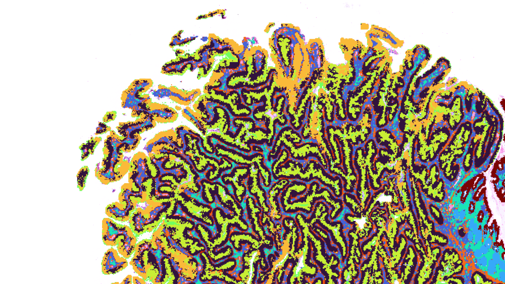
Visium Spatial
Assess spatial gene expression of FFPE or fresh frozen tissue sections. Harness whole transcriptome discovery while defining the relationship between cellular function and location.